


Osteoarthritis of the hip joint degenerative pathology, which is characterized by destruction of hyaline cartilage. The disease develops gradually, associated with pain and reduced range of motion. In the absence of medical intervention at the initial stages of osteoarthritis in a few years, there is atrophy of the thigh muscles. The injured limb is shortened, and the splicing of the joint space leads to partial or complete paralysis of the hip joint. Causes pathology become previous trauma, curvature of the spine, systemic diseases of musculoskeletal system.
Osteoarthritis usually is diagnosed in patients of middle age and older people. The diagnosis put on the basis of results of instrumental investigations — x-ray, MRI, CT, arthroscopy. Treatment of disease 1 and 2 degrees of severity conservative. In identifying ankylosis, or the ineffectiveness of drug therapy, surgical operation (arthrodesis, arthroplasty).
The mechanism of development of pathology
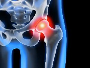
The hip joint consists of two bones — the iliac and femoral. The lower part of the Ilium presents her body, which participates in plexus with the femur, forming the upper section of the acetabulum. During the movement of the stationary glenoid fossa and the femoral head moves freely. This "hinge" device, the hip joint allows him to bend, stretch, rotate, promotes abstraction, bringing thighs. Free sliding of the articular structures provides a smooth, firm, elastic hyaline cartilage lining the socket and the femoral head. Its main function is the redistribution of loads during movement, prevent fast deterioration of bone tissue.
Under the influence of external or internal factors disturbed trophic cartilage. He doesn't have a circulatory system — nutrients fabric delivers synovial fluid. In case of arthrosis it thickens and becomes viscous. There is a deficiency of nutrients provokes a drying out of the surface of hyaline cartilage. It is covered with cracks, which leads to constant micro trauma to the tissues during flexion or extension of hip joint. The cartilages become thinner, lose their cushioning properties. To adapt to the increasing pressure, the bones are deformed. And against the background of deterioration of metabolism in the tissues of the progress of the destructive-degenerative changes.
Causes and predisposing factors
Idiopathic or primary osteoarthritis develops without any reason. It is believed that the destruction of cartilage occurs through the natural aging process, slowing the recovery process, decrease the production of collagen and other compounds necessary for proper regeneration of the structures of the hip joint. Secondary osteoarthritis occurs on a background of pre-existing pathologic condition. The most common causes of secondary disease include:
- previous injury — damage of the tendon unit, muscle tears, their complete separation from the bone base fractures, dislocations;
- developmental disorders of articulation, congenital dysplastic disorders;
- autoimmune pathology of rheumatoid, reactive, psoriatic arthritis, systemic lupus erythematosus;
- nonspecific inflammatory diseases, for example suppurative arthritis;
- specific infection, gonorrhea, syphilis, brucellosis, anaplasmosis, trichomoniasis, tuberculosis, osteomyelitis, encephalitis;
- disruption of the functioning of the endocrine system;
- degenerative pathology — osteochondropathy of the femoral head;
- hypermobility of the joints due to the production of "more loose" collagen what causes their excessive mobility, weakness of the ligaments.
Since the cause of development of osteoarthritis may be hemarthrosis (bleeding in the cavity of the hip joint), the precipitating factors include a violation of hematopoiesis. Prerequisites to the disease are excess weight, excessive exercise, sedentary lifestyle. To its development lead to a wrong organization of trainings, a deficiency in intake of foods with a high content of minerals, fat - and water-soluble vitamins. Postoperative arthrosis occurs several years after the surgical intervention, especially if it was accompanied by the removal of a large volume of tissue. Osteoarthritis of the hip joint cannot be transferred by inheritance. But in the presence of certain innate characteristics (metabolic disorders, skeleton structure) the probability of development is significantly increased.
Symptoms
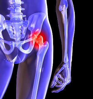
Leading symptoms of osteoarthritis of the hip joint pain while walking in hip and knee joint. A person suffers from stiffness in the limbs, stiffness, especially in the morning. To stabilize the joint, the patient begins to limp, changing his gait. Over time due to muscle atrophy and deformation of the joint the limb is noticeably shorter. Another characteristic feature of the pathology — limitation of hip abduction. For example, difficulties arise when trying to sit on a stool, legs apart to the sides.
Even "launched" ARTHRITIS can be cured at home! Just remember once a day to smear it...
For arthrosis of first degree characterized by periodic pain that occurs after intensive physical loads. They are located in the area of articulation and disappear after long periods of rest.
In the second degree osteoarthritis of the hip joints the pain intensity increases. Discomfort occur even at rest, extend to the hip and groin, worse when lifting weights or increasing physical activity. To address hip pain, a person begins with a scarcely perceptible limp. Marked limitation of motion of the joint, especially in abduction and internal rotation of the hip.
Arthrosis of the third degree is characterized by constant severe pain. When driving challenges arise, so when walking, the person is forced to use a cane or crutches. Due to the weakness of abductor muscles of the hip is offset from the pelvic bone in the frontal plane. To compensate for the events of shortening the patient's legs when the movement is tilted toward the affected limb. This provokes a shift of the center of gravity and increased stress on the joint. At this stage of the arthrosis develops a pronounced ankylosis of the joint.
| Degree | Radiographic signs |
| First | Changes are expressed not sharply. Joint gap narrowed moderately evenly, there is no destruction of the surface of the femur. On the outer or inner edge of the acetabulum, there was a slight bony growths |
| Second | The height of the joint space is significantly reduced due to its irregular intergrowths. Bone head of the femur is displaced upward, deformed, enlarged, its contours become uneven. Osteophytes are formed on the inner surface and the outer edge of the glenoid fossa |
| Third | There is a complete or partial fusion of the joint space. The head of the femur is strongly expanded. Multiple bone growths located on the surface of the acetabulum |
Diagnosis
When setting the diagnosis, the doctor considers the clinical manifestations of the disease, anamnesis, external examination of the patient and instrumental examinations. The most informative radiography. With its help, the estimated state of the hip joint, it sets the stage of its course, the degree of damage to cartilage tissues, and in some cases, the cause of development. If cervico-diaphyseal node is increased, and acetabulum bevelled and flattened, then with high probability it is possible to assume congenital dysplastic changes in the joints. Perthes disease indicates broken form of the thigh bone. X-ray allows the identification and preparation of post-traumatic osteoarthritis, despite lack of history of previous disease of injury. Also, use other diagnostic methods:
- CT helps to identify the expansion of the edges of the bony plates that formed osteophytes;
- MRI is done to evaluate the condition of the connective tissue structures and the extent of their involvement in the pathological process.
If necessary, the inner surface of the joint is examined with the help of arthroscopic instruments. Differential diagnosis done to rule out osteoarthritis, lumbar-sacral or thoracic degenerative disc disease. Pain in osteoarthritis may masquerade as clinical manifestations of radicular syndrome is caused by pinching or inflammation of a nerve. To exclude neurogenic pathology usually through a series of tests. Osteoarthritis of the hip joint necessarily differentialsa from the top of bursitis of the hip, ankylosing spondylitis, reactive arthritis. To exclude autoimmune pathologies are conducted biochemical studies of blood and synovial fluid.
Surgery
With the ineffectiveness of conservative therapy Il diagnostics of complicated pathology surgery performed. To restore cartilage in joints damaged by arthritis, no surgeries impossible, but with the right approach to treatment, compliance with all medical regulations, maintain proper lifestyle, medical gymnastics, regular courses of massage, vitamins and proper diet you can stop the process of destruction and destruction of cartilage and hip joints.